NSF-funded consortium will use X-ray lasers to study atomic structure of proteins, other molecules
Rice University will help probe the structure of ever-smaller molecules as part of a new National Science Foundation (NSF) consortium.
The NSF committed $25 million for a Science and Technology Center based at the University at Buffalo to explore the use of strong X-ray lasers to detail the atomic structures of molecules with a resolution approaching the nanoscale.

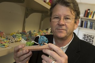
George Phillips will lead the Rice effort to probe the structure of molecules at the nanoscale as part of the new BioXFEL center, a Science and Technology Center funded by the National Science Foundation. Photo by Jeff Fitlow
Rice researchers led by George Phillips, the Ralph and Dorothy Looney Professor of Biochemistry and Cell Biology, expect to receive $1.25 million through the new BioXFEL center to create algorithms that sort out the flood of data provided by new X-ray laser crystallography techniques.
The ability to visualize protein is valuable to scientists who design drugs that interact with them. Research on viruses, for instance, benefits greatly from the ability to see how proteins are structured.
Phillips, a specialist in crystallography who returned to Rice from the University of Wisconsin, Madison, last year, said that while X-ray crystallography has come a long way since the technique was invented 100 years ago, a new class of lasers would take researchers deeper than ever into the structure of matter. It may also offer a new way to capture moving images of proteins.
“How you make the X-rays and how intense they are determine what you can do with them,” Phillips said. Current processes require targets like proteins to be frozen via crystallization, but proteins and other molecules are never static. “This is the next evolution,” he said.
The new technique will be based on an X-ray laser developed at Stanford University’s SLAC National Accelerator Laboratory that fires a pulse of light a billion times brighter than the best current sources.
“Physicists get their jollies slamming things into each other at high energies,” Phillips said. “Anytime you accelerate a charged particle, radiation is given off, and X-rays are generated like crazy, hitting the lead bricks and being wasted in these physics experiments.”
The realization that the light from accelerated particles could be used for its own sake led Stanford scientists to add a component to their accelerator to make really bright X-rays, he said. “This was a huge step forward because now we had access to incredibly intense X-ray beams,” Phillips said.
The beam has since been turned into an extremely accurate laser, one that can hit a target 1 micron across with a beam that lasts less than 100 femtoseconds. (A femtosecond is one-millionth of one-billionth of a second.)
The laser will let researchers gather data from streams of crystals, rather than one at a time, and may eventually allow them to piece together films that show how proteins move. As with current crystallography, the data will consist of captured diffraction patterns that mark the location of atoms in a crystal and allow researchers to determine its atomic structure.
“If we can parade molecules in front of the beam, maybe we can skip the crystallization part altogether,” he said.
Phillips said his immediate task will be to study variations in sets of data for molecules imaged by the laser. “I’m part of the team that will develop theory and methods for understanding the variations and propose algorithms for dealing with it,” he said. “At some point, I may put specimens in the beam for my own experimental program, but that’s not part of the immediate goal.”
Rice is one of eight institutions taking part in the consortium, along with Buffalo, the Hauptman-Woodward Institute, Cornell University, the University of Wisconsin at Milwaukee, Arizona State University, the University of California at San Francisco and Stanford University. The University of California at Davis will help with the creation and management of the center’s educational component.
“We’re very visual creatures,” Phillips said. “The cliché is ‘Seeing is believing.’ If you can see these atoms and molecules doing something, it’s very compelling. That’s what makes crystallography so popular, whether it’s for nanomaterials or biochemistry or chemistry or physics or whatever. It’s a way of visualizing things.”


Leave a Reply